Myasthenia gravis diagnosis and testing
 Fact-checked by
 Establishing a diagnosis of myasthenia gravis (MG), a rare neuromuscular disease, can be challenging because its symptoms are variable and may overlap with other disorders.
In MG, the immune system mistakenly attacks the sites where nerves and muscles communicate, causing symptoms of muscle weakness and fatigue that may affect various muscle groups.
The exact process of testing for MG can vary, but it usually starts with recognizing signs and symptoms in physical and neurological exams. Additional blood tests, evaluations of nerve, muscle, or breathing function, and imaging scans may then help establish the diagnosis and determine the MG type.
Signs that may lead to an MG diagnosis
While muscle weakness and fatigue can be caused by a number of disorders, the specific pattern of these symptoms may lead a doctor to suspect they’re due to MG. Specifically, doctors may consider MG if someone displays weakness in certain commonly affected muscle groups, such as the:
- eye (ocular) muscles: droopy eyelids (ptosis) and double vision (diplopia)
- head and neck (bulbar) muscles: difficulty chewing, swallowing, or speaking
- limb muscles: movement problems and changes in posture
- respiratory muscles: shortness of breath or difficulty breathing
Most people with MG initially experience eye muscle problems, with or without involvement of other muscles. About 15% of people have bulbar muscle involvement at symptom onset, and as many as about 25% of patients show signs of a myasthenic crisis, a severe breathing complication.
If the eyes are the only part of the body affected, it’s a sign of ocular MG. If more widespread weakness is occurring, it is a sign of generalized MG.
Regardless of what part of the body is affected, weakness in MG often fluctuates over time, typically worsening with activity and improving with rest. However, it is not usually accompanied by major neurological impairments, such as cognitive problems or issues with automatic bodily functions.
The first signs of MG might be recognized by a general practitioner or by an eye doctor through ptosis and diplopia testing. If someone meets one or more criteria on an MG symptoms checklist, doctors may recommend a thorough neurologist evaluation and follow-up testing.
Rapid assessment tests
Quick clinical tests can sometimes help support a doctor’s suspicion of MG. Their results may prompt further evaluation, but they can’t confirm a diagnosis of MG without other supporting evidence.
There are two rapid assessment tests that can evaluate whether ptosis might be related to MG:
- ice pack test
- Tensilon test
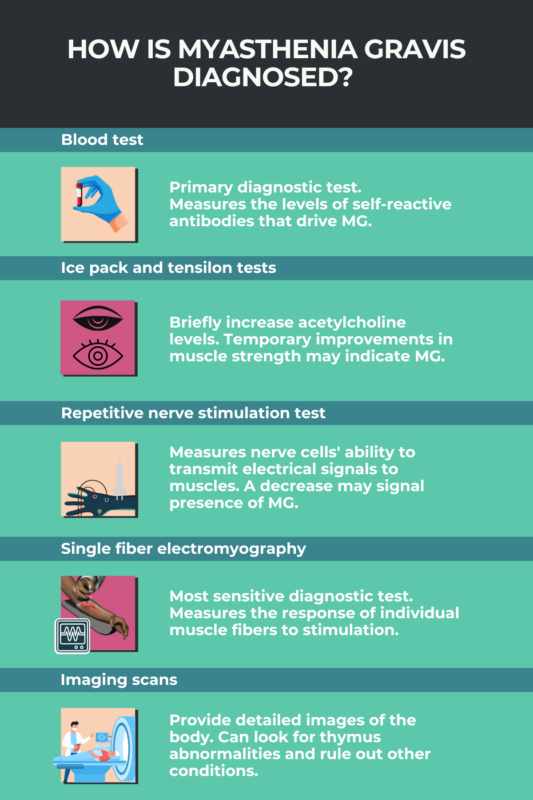
Ice pack test
The ice pack test for ptosis involves putting an ice pack on the eyelid for a few moments. A temporary reduction in ptosis after icing is consistent with MG, but not common if ptosis is related to a different underlying cause.
Scientists believe that this improvement occurs for several reasons, including because cold temperatures slow the breakdown of acetylcholine, the signaling molecule that triggers muscle contraction. Acetylcholine signaling is disrupted in MG, and increasing its availability by icing can enhance muscle contraction and reduce eyelid droopiness.
Tensilon test
The Tensilon test, also called the edrophonium test, involves injecting edrophonium chloride, a compound that blocks acetylcholine breakdown, to elicit an improvement in muscle strength. Similar to the ice pack test, the Tensilon test can temporarily relieve ptosis in someone with MG.
While this test was once common practice, clinicians now prefer the ice pack test because it is easier, cheaper, and safer. Endrophonium testing for MG is no longer approved in the U.S., but may be rarely used elsewhere.
Diagnostic tests
If there is enough evidence based on a person’s symptoms and initial clinical examinations, doctors may recommend additional diagnostic tests, including:

Living with MG
Essential Life Hacks for Myasthenia Gravis


Living with MG
Navigating Mental Health with Myasthenia Gravis
- blood tests: to identify self-targeting antibodies that cause MG
- repetitive nerve stimulation tests : to identify changes in nerve-muscle communication
- single-fiber electromyography: to test responses of individual muscle fibers
Blood test
Blood tests to identify the self-reactive antibodies that typically cause MG are the main way of diagnosing the condition. While there are no universally accepted MG diagnostic criteria, the presence of disease-causing antibodies, along with typical symptoms, is usually sufficient to support a diagnosis.
Antibodies targeting the acetylcholine receptor (AChR), a protein that helps muscle cells receive signals from nerve cells, are the most common cause of MG. About 80%-85% of people with generalized MG and 50%-70% with ocular MG have positive results on AChR antibody blood tests.
Blood tests can also detect antibodies against muscle-specific kinase (MuSK) and lipoprotein receptor-related protein 4 (LRP4), which are less common causes of MG. Both of these proteins help with the formation and function of neuromuscular junctions, where nerve cells and muscle cells meet to communicate.
If disease-causing antibodies aren’t detected but symptoms persist or worsen, doctors may recommend repeat testing, because some people who initially test negative later test positive.
In some cases, patients never test positive for antibodies. This is called seronegative MG. These individuals may require additional nerve and muscle tests to confirm the diagnosis.
Repetitive nerve stimulation test
A repetitive nerve stimulation (RNS) test allows doctors to measure how well nerve cells transmit signals to muscles, and how muscles respond.
During the test, an electrode is placed on the skin and repeatedly stimulates a nerve near a muscle of interest. Normally, a muscle would have a strong response throughout repeated stimulation.
In people with MG, however, the muscle response becomes weaker over time, reflecting how symptoms often worsen with fatigue or activity. Abnormal RNS results are more common in generalized MG than ocular MG.
Single fiber electromyography
While RNS measures the response of a whole muscle, single fiber electromyography (EMG) measures the activity of single muscle fibers, making it a more sensitive test for diagnosing MG.
A fine wire electrode is inserted through the skin into a muscle — usually in the forehead above the eyebrow — where it measures the activity of nearby muscle fibers. Normally, individual muscle fibers controlled by the same nerve should fire at the same time when the nerve sends a signal. But in MG, this timing may be more variable.
This can help capture subtle changes in muscle activity that can’t be detected elsewhere, making the test useful for reaching an ocular MG diagnosis and for identifying mild cases without other diagnostic findings. However, other disorders involving the neuromuscular junction can yield similar findings, so further tests may be needed to exclude them.
Imaging and respiratory assessments
Other tests may play a supporting role in reaching an MG diagnosis. These include:
- MRI or CT scans of the thymus gland to detect abnormalities that contribute to MG
- pulmonary function tests to assess respiratory muscle weakness
Imaging scans
Imaging scans, most commonly CTs or MRIs, may help clinicians identify problems in the thymus gland, an immune organ in the chest. Thymus abnormalities — including thymus enlargement, called thymic hyperplasia, or tumors, known as thymomas — are thought to contribute to the development of MG-causing antibodies in many cases.
Imaging scans can also be used to rule out other neurological disorders with overlapping symptoms or to identify possible causes of ocular symptoms.
Pulmonary function tests
Pulmonary function tests (PFTs) can identify problems with the muscles that support breathing.
They’re often used after an MG diagnosis to predict a person’s risk of serious respiratory complications. However, some people with severe forms of MG, particularly those with MuSK antibodies, show respiratory symptoms early in the disease course, so PFTs may also be used during the diagnostic process.
The most common PFTs use a small device called a spirometer to measure how much air a person can inhale and exhale.
Ruling out similar conditions
Symptoms of MG can resemble those of several other conditions, including other neuromuscular junction disorders. Doctors must rule out these other possible causes of symptoms to confirm the MG diagnosis.
Conditions that may be considered in the differential diagnosis of MG include:
- Lambert-Eaton myasthenic syndrome (LEMS): an autoimmune disorder that affects the neuromuscular junction
- amyotrophic lateral sclerosis (ALS): a neurodegenerative disease caused by the death of nerve cells that support movement
- botulism: an illness caused by nerve toxins, often contracted from improperly canned foods or deep wounds
- thyroid eye disease: an autoimmune disease that affects the tissues behind the eyes
- oculopharyngeal muscular dystrophy (OPMD): a genetic disorder that affects muscles of the eyes and throat
- Guillain-Barré syndrome: an autoimmune disease that affects nerves outside the brain and spinal cord
Although there is overlap between these conditions and MG, there are also clinical differences that help doctors differentiate them:
| Conditions | Similarities to MG | Differences from MG |
|---|---|---|
| LEMS | General muscle weakness, fatigue | Ocular and bulbar muscle weakness are less prominent, other neurological symptoms may be present, and different disease-causing antibodies |
| ALS | Bulbar and general muscle weakness | Ocular muscle weakness is typically less prominent, other motor symptoms are present, symptoms are progressive and do not fluctuate |
| Botulism | Ocular, bulbar, and general muscle weakness | Begins suddenly, causes paralysis and pupil dilation, other neurological symptoms may be prominent |
| Thyroid eye disease | Double vision | Droopy eyelids are uncommon, but other eye-related symptoms are present |
| OPMD | Double vision, droopy eyelids, difficulty swallowing | Limb weakness is typically less prominent, develops and progresses slowly |
| Guillain-Barré Syndrome | General muscle weakness | Nerve pain and other sensory symptoms may be present, muscle reflexes may be altered |
Next steps after diagnosis
After an MG diagnosis, patients will work with their doctors to develop a comprehensive treatment plan to suit their individual needs. This may involve:
- medications
- surgery to remove the thymus gland (thymectomy)
- lifestyle changes, such as diet adjustments to cope with swallowing problems or exercise regimens to maintain muscle strength
Patients should also work with their care team to track and avoid disease triggers, such as heat, stress, or medications, that could make their symptoms worse.
Myasthenia Gravis News is strictly a news and information website about the disease. It does not provide medical advice, diagnosis, or treatment. This content is not intended to be a substitute for professional medical advice, diagnosis, or treatment. Always seek the advice of your physician or other qualified health provider with any questions you may have regarding a medical condition. Never disregard professional medical advice or delay in seeking it because of something you have read on this website.