Fact-checked by
 Ocular myasthenia gravis (MG) is a specific form of the autoimmune condition myasthenia gravis in which only the muscles that control the eyes and eyelids are affected.
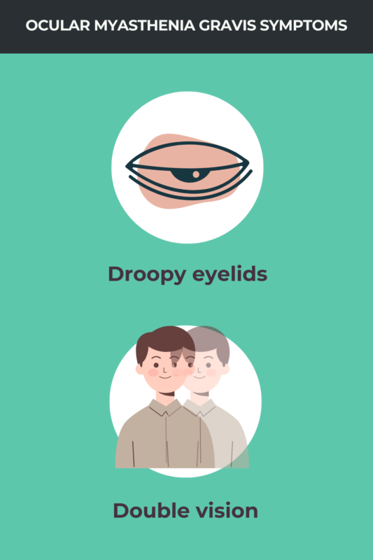
In people with MG, the immune system mistakenly attacks the proteins that allow nerves and muscles to communicate. When these signals are interrupted, the muscles become easily tired and weak. For many patients, this process begins in the eyes, frequently resulting in drooping eyelids or double vision.
When these symptoms stay confined to the eye area without spreading to the rest of the body, the condition is classified as ocular myasthenia gravis. While some patients will only ever experience eye-related issues, about 20% to 60% of people with ocular MG will eventually progress to generalized MG, where muscle weakness becomes more widespread throughout the body.
Causes
Abnormal antibodies cause MG by disrupting the communication between nerves and muscles. In about half of all cases, these antibodies target the acetylcholine receptor (AChR), a protein that transmits signals from nerve cells to muscles. When these receptors are blocked, muscles cannot get the message to contract. Other types of antibodies can also damage the neuromuscular junction, the specific site where these signals are passed from nerve to muscle.
In some cases, a person may have all the classic symptoms of the condition but no detectable antibodies in their blood. This is known as seronegative MG. Scientists are working to understand exactly what causes the disease to develop in these individuals.
Researchers are also investigating why the eyes are so uniquely vulnerable. Several factors may explain why eye muscles are often the first or only ones affected:
- High activity levels: Eye movements require constant, rapid-fire signals from nerve cells, making them more likely to show fatigue when communication is disrupted.
- Lower signal strength: Nerve cells in the eye release smaller amounts of the molecules needed to trigger muscle contractions compared to other parts of the body.
- Fewer receptors: Eye muscles naturally have fewer AChR proteins available to receive incoming signals.
- Unique architecture: The physical structure of the neuromuscular junction in the eyes is different from that found in larger limb muscles.
Common symptoms
The symptoms of ocular MG are restricted to the eyes. Common MG eye symptoms include:
- double vision (diplopia): This occurs when a person sees two images of a single object.
- drooping eyelids (ptosis): This can affect one or both eyes and may make it difficult to keep the eyes open.
While these symptoms can cause visual disturbances and make it hard to focus, they do not usually damage the eyes directly or lead to permanent blindness.
A key feature of this condition is fluctuating muscle weakness that worsens with fatigue. In ocular MG, this means that drooping eyelids often improve after a period of rest but become more noticeable after repeated use or at the end of a long day. Doctors specifically look for these shifting patterns to help diagnose ocular MG and distinguish it from other eye-related conditions.
Testing & diagnosis
Confirming an ocular MG diagnosis typically requires a combination of clinical exams and specialized tests. This thorough process helps doctors rule out other potential causes of eye issues, including other autoimmune ocular disorders.
Because the condition is specialized, a neurologist, ophthalmologist, or a neuro-ophthalmologist — a specialist who focuses on the relationship between the eyes and the nervous system — will perform a detailed evaluation. During this exam, they will assess eyelid movement, assess changes in muscle strength over time, and review the patient’s history of eye symptoms.
Quick bedside tests
If a doctor suspects that your symptoms are caused by ocular MG, they may perform a series of quick bedside exams. While these tests cannot confirm a diagnosis on their own, they help strengthen clinical suspicion and determine which specialized tests are needed next.
Common bedside evaluations include:
- Cogan’s lid twitch: This test identifies eyelid movements characteristic of MG. A doctor will ask you to look downward for several seconds and then return your gaze to the center. People with the condition often show a brief, twitch-like overshooting of the upper eyelid as it moves back up, which is caused by muscle fatigue and recovery.
- ice pack test: This is used to determine whether cold temperatures improve droopy eyelids. A doctor briefly places an ice pack over a closed eye; if the drooping eyelid is temporarily eased, it is a strong indicator of ocular MG. Many clinics prefer this method because it is simple, inexpensive, and carries very few side effects.
- Tensilon test: This involves an injection of edrophonium chloride. Like the ice pack test, it is designed to see if muscle strength increases temporarily. If the drooping improves immediately after the injection, it suggests the nerve-muscle communication is being affected by MG.
Beyond these specific exams, doctors may also evaluate fluctuating muscle weakness by checking for eyelid drooping after a period of rest, rapid blinking, or by asking you to squeeze your eyes shut as tightly as possible. These simple physical challenges help reveal the unique way MG fatigue affects the small muscles of the eye.
Blood work & SFEMG
Additional diagnostic tests can provide the definitive evidence needed for an ocular MG diagnosis:
- Blood tests: These are used to identify the specific antibodies that interfere with nerve-muscle communication. While finding these antibodies can confirm the condition, many people with this form of the disease are seronegative, meaning their blood tests may come back negative despite having symptoms.
- Single-fiber EMG: Because blood tests aren’t always conclusive, doctors often rely on single-fiber EMG, which is considered the most sensitive diagnostic tool for ocular MG.
During a single-fiber EMG, a specialist inserts a very thin needle electrode into a muscle to record the electrical activity of individual muscle fibers. The test looks for “jitter,” which refers to subtle delays or inconsistencies in the timing of signals between the nerve and the muscle.
By detecting these tiny signaling errors, the test can help doctors identify the disease even when other evaluations have failed to provide a clear answer.
Treatment options
Treatment for ocular MG focuses on relieving eye-related symptoms and improving quality of life. Because every patient experiences the condition differently, doctors tailor the approach using several different options:
- Approved MG medications: These often include acetylcholinesterase inhibitors, which help strengthen the signals between nerves and muscles to improve eyelid control and focus.
- Immune-suppressing therapies: If standard medications aren’t enough, doctors may prescribe steroids or other drugs to dampen the immune system’s mistaken attack on the body.
- Vision aids: Simple tools like prisms in eyeglasses can help correct double vision, while eyelid tape or specialized “ptosis crutches” attached to glasses can help keep drooping eyelids open.
- Surgical interventions: In stable cases where symptoms don’t fluctuate, eyelid or eye muscle surgery can sometimes provide a more permanent correction for drooping or misalignment.
Another option is the removal of the thymus gland, a procedure known as a thymectomy. The thymus is an immune organ that scientists believe plays a key role in producing the abnormal antibodies that cause MG.
While a thymectomy is a standard treatment for many people with generalized MG, its effectiveness for those with the purely ocular form is still being studied. However, the procedure is often strongly recommended if a patient is found to have a thymoma (a tumor on the thymus) or if they are at high risk of the disease spreading to other muscles in the body.
Medications
Several types of medications can help manage the symptoms of ocular MG by improving muscle strength or calming the immune system:
- Acetylcholinesterase inhibitors: Medications such as Mestinon (pyridostigmine bromide) are often the first-line treatment. They work by increasing the amount of signaling chemicals available at the neuromuscular junction, making it easier for muscles to contract.
- Corticosteroids: These anti-inflammatory drugs help suppress the overactive immune response. They are frequently used to reduce drooping and double vision when other medications aren’t enough.
- General immunosuppressants: If steroids cause too many side effects or aren’t effective long-term, doctors may use other medications to dampen the immune system’s attack on muscle receptors.
In recent years, scientists have developed several biologic therapies approved for generalized MG. These include complement inhibitors and neonatal Fc receptor (FcRn) blockers, which target specific parts of the immune system responsible for the disease. While these are currently used for more widespread weakness, researchers and clinicians are investigating whether they can provide the same relief for those with symptoms limited to the eyes.
Ongoing clinical trials are focused on a major goal: determining whether new or existing treatments can prevent the disease from progressing. By intervening early, scientists hope to find ways to keep the condition localized to the eyes and prevent it from progressing to more severe, generalized muscle weakness.
Vision aids
In addition to medication, several non-drug interventions can help manage vision challenges. These options focus on improving daily function and comfort:
- Vision aids: To reduce double vision, doctors may suggest using eye patches to block one image or wearing special prism eyeglasses. Prisms work by bending light before it enters the eye, helping to realign the two separate images into a single, clear one.
- Ptosis crutches: If a drooping eyelid makes it difficult to see, a small attachment called an eyelid crutch can be added to the frame of your glasses. These crutches physically hold the eyelid open. While effective at clearing the field of vision, they can sometimes cause eye irritation or dryness since the eye may not blink as naturally.
- Eyelid surgery: If symptoms are stable and do not respond to medication, doctors may recommend a surgical procedure to lift the eyelid. This is generally considered only when the drooping significantly interferes with vision and other treatments have not provided enough relief.
Prognosis
Most people living with ocular MG have a positive long-term outlook when their symptoms are managed effectively. The condition generally does not shorten a person’s life expectancy, though persistent vision problems can make daily tasks like reading, working on a computer, or driving more challenging.
For many, the primary concern is whether the weakness will spread. Some individuals will eventually develop generalized MG, which involves limb weakness or bulbar symptoms. These symptoms, such as difficulty swallowing or slurred speech, indicate that the muscles in the face, mouth, and throat have become weak.
This progression most commonly occurs within the first two years after the onset of initial eye symptoms. If the weakness remains confined to the eyes beyond this two-year window, there is a relatively low generalization risk, and the condition is more likely to remain ocular.
Several factors may increase the likelihood of the disease becoming generalized, including:
- antibody status: Testing positive for AChR antibodies, especially at higher levels
- thymus health: Discovering abnormalities or tumors in the thymus gland
- age: Being older than 50 at the time of diagnosis
- symptom severity: Experiencing more intense or persistent eye-related issues
- diagnostic results: Showing significant abnormalities on specialized nerve and muscle signaling tests
Understanding these risks helps doctors monitor the condition closely and adjust treatment plans to provide the best possible support for each patient.
Myasthenia Gravis News is strictly a news and information website about the disease. It does not provide medical advice, diagnosis, or treatment. This content is not intended to be a substitute for professional medical advice, diagnosis, or treatment. Always seek the advice of your physician or other qualified health provider with any questions you may have regarding a medical condition. Never disregard professional medical advice or delay in seeking it because of something you have read on this website.