
Diverse mechanisms found for myasthenia gravis’ AChR antibodies
Despite variations, all the antibodies interfered with AChR protein activation
Written by |
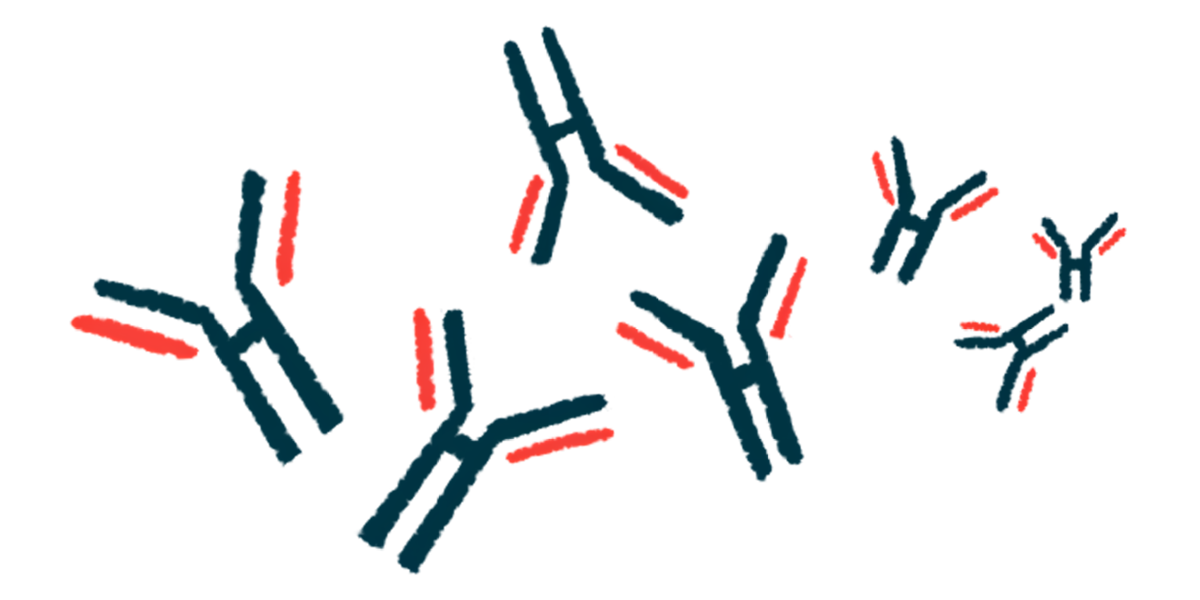
Disease-causing antibodies from different myasthenia gravis (MG) patients bind to and affect the function of acetylcholine receptors (AChRs) in different ways, a study finds.
While the mechanism varied, all the antibodies ultimately interfered with the usual activation of AChRs, which are the proteins important for normal muscle contractions commonly targeted in the autoimmune processes that drive MG.
“We were able to visualize, for the first time, how individual myasthenia gravis antibodies engage the receptor and interfere with its function,” Colleen Noviello, PhD, a project scientist at the University of California (UC), San Diego and one of the study’s senior authors, said in a university news story. “These insights offer a new path forward for designing treatments that can precisely target the disease-causing mechanisms in each patient.”
The study, “Autoimmune mechanisms elucidated through muscle acetylcholine receptor structures,” was published in Cell.
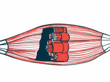
Muscle contractions that enable voluntary movement occur when motor nerve cells release a signaling molecule called acetylcholine at the neuromuscular junction (NMJ), the site where nerve and muscle cells meet and communicate, that binds to AChRs residing on muscle cells at the other side of the junction. AChRs are protein channels that open when bound to acetylcholine, allowing a flow of ions that cause muscle cells to contract.
In MG, the immune system mistakenly produces self-reactive antibodies, or autoantibodies, that target NMJ proteins, most often AChRs, that impede this process, resulting in muscle weakness and fatigue.
Various treatments are available for MG, including boosting acetylcholine levels, suppressing the immune system, or facilitating the breakdown of the autoantibodies. Responses to different therapeutic approaches vary, however, which “suggests that the underlying [disease-driving] mechanisms differ among patients,” the researchers wrote.

Diversity in antibodies’ role in MG
Characterizing exactly how different antibodies might interact with AChRs in people has been a challenge, in part because techniques to visualize the detailed structure of these receptors have been limited. To overcome this, the scientists forced the production of human muscle AChRs in cells grown in the lab and then applied an advanced imaging technique called cryoelectron microscopy to visualize the three-dimensional structure.
They then looked at how MG-causing autoantibodies from six patients interacted with the receptors. These antibodies appeared to bind to AChRs in different places, affecting their function in distinct ways. For example, some directly blocked or destabilized the ability of acetylcholine to bind to AChRs, while others influenced the channels’ ability to allow typical ion flow.
We revealed a surprising diversity in how autoantibodies contribute to myasthenia gravis. This knowledge helps explain why some patients respond differently to treatments and provides a foundation for developing more personalized therapies.
Other processes were also sometimes triggered, such as those that caused AChRs to be internalized off the cell surface or that activated immune pathways that could destroy AChRs. A combination of different mechanisms was also seen.
All the autoantibodies disrupted normal AChR function, irrespective of how they did it, however.
“We revealed a surprising diversity in how autoantibodies contribute to myasthenia gravis,” said Ryan Hibbs, PhD, UC San Diego professor and one of the study’s senior authors. “This knowledge helps explain why some patients respond differently to treatments and provides a foundation for developing more personalized therapies.”
Developing therapies could consider these diverse mechanisms, rather than taking a broader immunosuppressive approach, according to the researchers, who said that approach may also be relevant for other conditions driven by self-reactive antibodies.
“This study not only advances our understanding of myasthenia gravis, but also sheds light on other autoimmune diseases … offering hope for more precise and effective treatment strategies,” Hibbs said.
The study was funded by the American Heart Association, the National Institutes of Health, and the Myasthenia Gravis Foundation of America.


Leave a comment
Fill in the required fields to post. Your email address will not be published.