
Test That Tracks Eye Movements May Help in Diagnosing MG
Written by |
Videonystagmography, a video-tracking system used to investigate balance disorders, can also detect eye muscle fatigue and recovery in myasthenia gravis (MG) patients, making it a potentially useful diagnostic test for MG, a study reported.
The study, “A Novel Diagnostic Method for Myasthenia Gravis,” was published in the journal Muscle and Nerve.
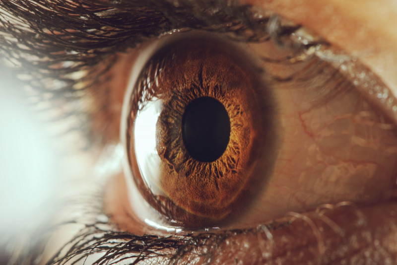
Muscle weakness characterizes MG, due to damage at the neuromuscular junction where nerve and muscle cells communicate. This weakness can be more generalized or more restricted to specific muscle groups, like those controlling eye and eyelid movements — a disease subtype known as ocular MG.
While a combination of clinical and symptom history, blood tests, and nerve stimulation tests are used to diagnose MG, diagnosing ocular MG can be more challenging.
Researchers in Turkey assessed the potential of videonystagmography (VNG) in detecting drops in eye muscle activity, and its usefulness a possible diagnostic test for MG.
VNG uses specialized infrared cameras to track rapid and involuntary eye movements known as nystagmus. Here, researchers used VNG to track eye movements in people with myasthenia gravis — each eye individually for 60 seconds — while the person followed a moving target. Changes in eye movement speed were used as indicators of muscle fatigue.
Their analysis included 34 MG patients — 13 with predominantly ocular MG and 21 generalized MG — treated at an Istanbul clinic between July 2017 and December 2019, and 23 healthy participants who served as controls. Patients were either newly diagnosed or new to immunomodulatory therapy.
Self-reactive antibodies directed against the acetylcholine receptor, one of the main causes of MG, were detected in 24 (71%) patients, while antibodies targeting muscle-specific kinase (MuSK) were found in two (6%) others. None of these two self-reactive antibodies were evident in the blood of eight (23%) patients.
All patients had ptosis (droopy eyelids) and 16 also had diplopia (double vision).
Their mean MG composite score, a measure of signs and symptoms that ranges from zero to 50, was 8.8, and one patient had problems chewing solid food.
Test results showed a slowing in the movement speed of each eye in MG patients relative to controls, which was detected several times during the 60-second recordings.
Investigators then assessed and compared three parameters among patients, and between patients and controls. These were maximum gain — the highest speed gain — that usually occurs at the beginning of the test; minimum gain, corresponding to the lowest speed gain, the result of fatigue caused by eye movements; and peak gain, corresponding to the average speed gain.
The mean maximum gain in the right eye was similar in patients with ocular and generalized MG (1.18 vs. 1.27), but lower in the left eye of those with generalized MG (1.36 vs. 1.14). In fact, the mean maximum gain for the left eye was higher among ocular MG patients compared with controls (1.36 vs. 1.17).
For both eyes, the mean minimum gain was significantly lower among MG patients compared with controls, although not between the two patient groups. Analyses also showed that MG patients experienced a mean drop of 90% in eye movement gain, compared with a mean drop of 17% among controls.
Researchers estimated that a movement gain drop of 48% in the right eye, 41% in the left eye, and 24% in both eyes could be used as threshold values to distinguish MG patients from healthy people with 100% sensitivity and specificity. (Sensitivity is a test’s ability to correctly identify those with a disease, while specificity refers to its ability to correctly identify those without it.)
After reaching minimum velocity, ocular MG patients had a mean gain of 1.54 seconds in eye movement speed, and those with generalized disease a mean gain of 1.13 seconds. No patterns of gain reduction or increment were evident among controls.
“This study shows that recording eye movement with VNG offers a quantifiable method of documenting [extraocular muscle] activities and effectively demonstrates muscle fatigue and recovery,” the researchers wrote.
The drop in extraocular muscle movement recorded in patients using VNG also ” begins to improve within one to two seconds after reaching minimum velocity, analogous to traditional low-frequency repetitive nerve stimulation testing and its U- shaped pattern,” they added.
“Thus, VNG may be a promising diagnostic test for MG,” the team concluded.


Leave a comment
Fill in the required fields to post. Your email address will not be published.