
Modified Method for Thymus Removal Can Offer MG Patients Less Scarring, Case Report Shows
Written by |
A new, modified method for removal of the thymus (thymectomy) in patients with myasthenia gravis can help achieve complete resection along with minimal scarring, leading to better cosmetic results, a new case report shows.
The study, “Modified unilateral video-assisted thoracoscopic extended thymectomy for myasthenia gravis using 5-mm incisions: A case report,” was published in the journal Medicine.
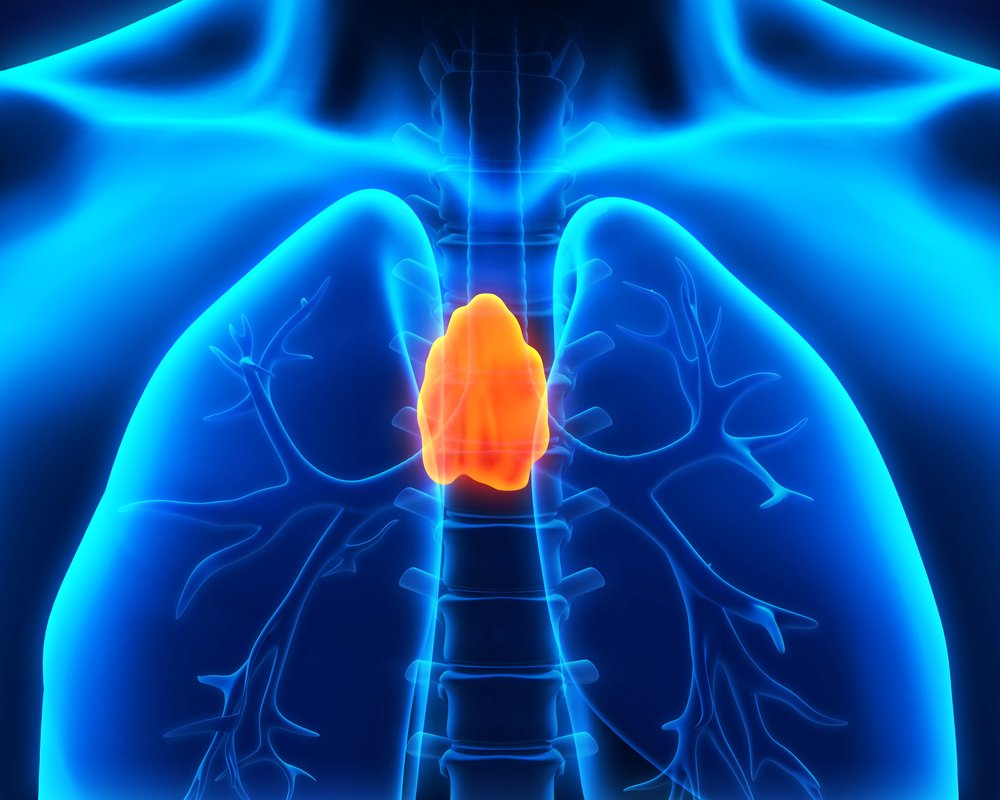
Myasthenia gravis (MG) is a chronic autoimmune disease responsible for weakness in skeletal muscles. Thymectomy (a procedure to remove the thymus) is an established therapy for treating MG.
A bilateral video-assisted thoracoscopic extended thymectomy (VATET), conducted using an endoscope, has been commonly used to treat MG and has several advantages over a traditional trans-sternal thymectomy. These include less tissue injury, reduced time in the hospital, and a better cosmetic result.
Unilateral VATET is an alternative approach to a bilateral VATET that has comparable long-term outcomes but reduces operating time.
In this case report, physicians describe the case of a 22-year-old Chinese woman with a six-year history of Mmyasthenia gravis who was admitted to a hospital with bilateral ptosis (drooping eyelids), diplopia (double vision), and intermittent dysphagia (difficulty swallowing).
A computed tomography (CT) scan of the patient revealed thymic hyperplasia — an enlarged thymus.
To treat the woman’s hyperplasia, physicians developed a minimally invasive technique that can help remove the thymus and adjacent fatty tissue using a 5 mm camera port.
While a thymus that is less than 4 centimeters in diameter can be removed as a complete mass through the port, this is often not the case for larger specimens.
To overcome this issue, physicians used scissors to shred the thymus within a specimen bag (introduced via the camera port) before its removal.
Thymectomy can often lead to scarring. In this case, scarring was kept to a minimum because of the use of the small 5 mm port.
Another advantage of the smaller port is that it exerts less pressure on the intercostal nerves (nerves connected to the spine) and can potentially result in less postoperative pain, including chronic pain.
The surgery was performed without complications and with very little bleeding. The patient had a chest X-ray a day after the procedure to confirm there were no post-operative complications.
The woman was discharged three days after surgery and was prescribed pyridostigmine bromide, a common medication for MG used to improve muscle strength.
Three months after surgery, the woman’s MG symptoms were well-controlled and her muscle strength returned to normal. A CT scan showed no residual thymus tissue. She stopped taking pyridostigmine bromide six months after the procedure.
“This modified VATET method could be a useful approach to the management of non-thymomatous MG. The ability to achieve complete dissection with good cosmetic results may lead to wider acceptance of this technique by patients with MG and their neurologists for earlier thymectomy and improved outcomes,” the authors said in the case study.


Jose Armando Cano Mata, from Monterrey, Mexico
I want to know, if,Calcium, Magnesium,Zinc, could help the syptom in M.G., I have 15 years with M:G:,my surgery of thymus, in the first two years after that, don't result how My Doctor expected,I have taken Mestinon,(4.0 pills at day or 60 mg) and one a day Myfortic,360 mg,since 8 years ago, each 4 months I recived Octagam or medicine like this