Low Frequency of Certain Immune Cells May Help Identify MG
Written by |

Lower frequency of a specific subset of immune cells in the bloodstream may help identify myasthenia gravis (MG) in cases difficult to diagnose, specifically those testing negative for MG autoantibodies, a study suggests.
Published in the journal Muscle & Nerve, the study, “Reduced plasmablast frequency is associated with seronegative myasthenia gravis,” was led by scientists at Duke University in North Carolina.
The majority (90%) of MG patients test positive for self-reactive antibodies, or autoantibodies, that target acetylcholine receptors (AChRs), muscle-specific tyrosine kinase, or lipoprotein receptor-related protein 4, which impair the communication between nerves and muscles.
The remaining patients are seronegative (negative in a blood test), since no such autoantibodies are detected. In these cases, MG is largely diagnosed using other tests, such as repetitive nerve stimulation (RNS) and single-fiber electromyography (SFEMG), among others.
These tests measure a nerve’s ability to transmit a signal to the muscle (RNS) and whether muscle fibers respond to repeated electrical stimulation (SFEMG).
Such tests, however, are “time consuming, require special training, and can be impractical in some clinical practice settings that do not evaluate a large number of MG patients,” the researchers wrote. “Alternative diagnostic approaches for seronegative MG are needed.”
Also, because it is more challenging to diagnose, the mechanisms underlying seronegative MG remain poorly understood.
Researchers at Duke and their colleagues evaluated the levels of immune cells in blood samples of people with seronegative MG. This analysis, besides shedding light on the disease mechanisms, also was intended to identify biomarkers that help diagnose seronegative MG.
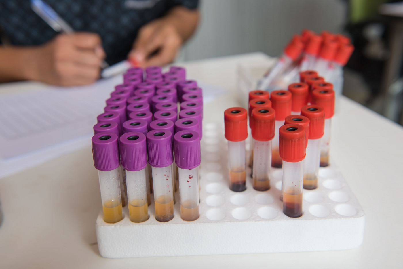
In total, they profiled blood samples from 95 MG patients, including 68 with seronegative MG and 27 patients positive for anti-AChRs autoantibodies, along with 46 healthy individuals who served as controls.
The team compared the levels of 12 immune cell subgroups between the controls and the MG patients.
A first analysis (the discovery phase) included 47 seronegative MG (median age 61.5 years) and 25 controls who were younger — median age 38. Few MG patients had undergone surgical removal of the thymus, or thymectomy, and below half were taking immunosuppressive therapies.
Results of the discovery phase revealed that lower frequency of a subgroup of immune B-cells — having the CD19 marker, not having CD20 and with high levels of the CD38 protein — was linked significantly with a higher probability of having seronegative MG, when compared to healthy controls.
These results were confirmed in the replication phase, which included 21 seronegative MG patients and 21 controls. In an exploratory analysis, increased frequency of such cells was associated with higher odds of being positive for anti-AChR antibodies than having seronegative MG. Notably, patients with anti-AChRs antibodies in this analysis were older and had higher disease severity than the seronegative MG participants.
Patients with AChRs-positive MG had increased frequency of interleukin (IL)-21 producing immune cells, called helper T-cells, with these cells associating with a diagnosis of AChRs-positive MG when compared to seronegative MG.
Overall, these results suggest that a reduction in the frequency of a specific subset of immune cells is “strongly associated with a SN [seronegative] MG diagnosis and may be a useful diagnostic biomarker in the future,” the researchers wrote.


Leave a comment
Fill in the required fields to post. Your email address will not be published.