Finding of Myasthenia Gravis After Surgical Removal of Thymus May Signal Cancer’s Return, Case Study Finds


Between 30 percent and 50 percent of all thymoma patients – people treated for a tumor in the thymus gland – go on to develop myasthenia gravis, and as a case study reports, this disease’s appearance can sometimes be associated with a return of the cancer.
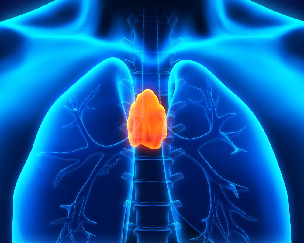
The study detailed an older woman who developed myasthenia gravis four months after total thymectomy, or surgical removal of the thymus gland. This gland is a lymphoid organ that produces T-cells, a type of white blood cell of importance to the immune system.
“Myasthenia gravis appearing after thymectomy heralding recurrent thymoma” was published in the journal Acta Chirurgica Belgica.
The 72-year-old woman went to the hospital after feeling progressive fatigue and shortness of breath during minimal exercise. Clinical examination of her chest using computed tomography revealed a tumor adherent to the right atrium of the mediastinum — the chest cavity between the lungs that contains the heart, esophagus, trachea, among others.
The patient underwent a total thymectomy – the preferred treatment for these patients because of the cancer’s malignant potential.
Four months later, she began to complain of difficulties swallowing and fatigue during exercise. After eight months of progressive complaints, clinicians diagnosed her with myasthenia gravis after blood tests revealed high levels of acetylcholine receptor-antibodies (18.01 nmol/L), a marker for the disease.
Local recurrence of the thymoma was found via positron emission tomographic (PET) scanning. This imaging technique uses small amounts of radioactive materials – called radiotracers – to evaluate organs and tissues.
The radiotracer is first injected intravenously (directly into the blood). After that, the PET scanner moves slowly over the part of the body being examined, where it detects the subatomic particles emitted by the radiotracer. A computer analyses this data and converts it into images inside the body – the amount of the radiotracer that accumulates in tissues allows clinicians to assess how well organs and tissues are functioning.
The nodule was surgically removed and the patient’s myasthenia gravis was treated with Mestinon (pyridostigmine) and a corticosteroid – a suppressor of the immune system – called Medrol (methylprednisolone). She refused radiotherapy.
Overall and in agreement with previous studies, this case report concluded that “repeat thymectomy should be attempted for patients with refractory MG [difficult to treat myasthenia gravis] after a previous thymectomy. Though complete remissions are rare, 60–70% cases report clinical improvement.”