Th22/Tc22 Cells May Be Involved in Development of Myasthenia Gravis


Two subtypes of immune cells, Th22 and Tc22, may be directly involved in the development of myasthenia gravis.
The study with that finding, “High Frequency of Tc22 and Th22 Cells in Myasthenia Gravis Patients and Their Significant Reduction after Thymectomy,” was published in Neuroimmunomodulation.
Myasthenia gravis (MG) is an autoimmune disease caused by the abnormal production of antibodies against acetylcholine receptors that are necessary for muscle contraction. The disorder is often accompanied by thymus (the gland that produces T-cells) anomalies, such as gland enlargement or hyperplasia (65% of the cases), and tumors or thymoma (10-15% of the cases).
For most patients experiencing these symptoms, a thymectomy (surgical removal of the thymus, TE) often is recommended as a therapeutic approach to stop B-cells in the thymus from producing autoantibodies that mistakenly target and attack the connections between nerve and muscle cells in MG patients.
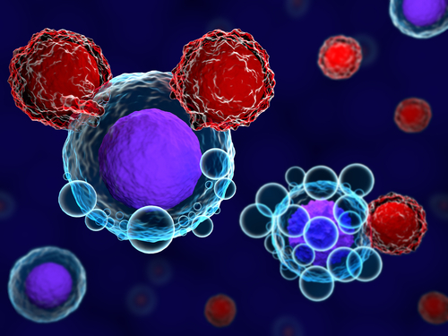
B cells are part of the immune system and produce antibodies against specific substances seen as threats (antigens). T helper cells (a subtype of T-cells that regulate the function of B-cells and killer T-cells) have been shown to further induce the production of these harmful autoantibodies.
Although the role of the thymus in MG autoimmunity has been described previously, it also is known that this gland plays a major role in the selection and removal of self-reactive T-cells, meaning T-cells that were wrongly instructed to target and destroy other cells or molecules that should not be perceived as a threat.
In addition, many studies have shown that Th22 and Tc22, a sub-group of T-helper cells, are involved in the development of several autoimmune diseases.
Researchers collected blood samples and isolated peripheral blood mononuclear cells (PBMCs) — a general term that comprises all immune cells with a round nucleus — from 12 healthy control subjects and 12 MG patients, six months before and after TE. Their aim was to assess whether the levels of Th22 and Tc22 cells are indeed altered in patients diagnosed with MG before and after TE
The mean percentage of Tc22 cells in MG patients before TE (0.97%) was significantly higher in comparison with healthy control subjects (0.49%). However, after TE, the levels of Tc22 cells in MG patients decreased substantially (0.69%).
Unlike Tc22 cells, the mean percentage of Th22 cells in MG patients before TE (1.75%) was identical to healthy controls, but after TE the levels of Th22 cells also dropped significantly (1.09%).
Although the precise role of T-helper cells still warrants further research, these findings suggest that Th22 and Tc22 cells could actively participate in the development of MG.
“To the best of our knowledge, this paper is the first to study Th22 and Tc22 cells in MG patients before and after TE,” the authors wrote.
“However, our study was limited by a small study population, short-term follow-up of patients (6 months), the absence of a pathophysiologic investigation of T cell transcription factors, and no data regarding cytokines. Larger scale studies are required to further clarify the role of these cells in MG pathogenesis [disease development],” the researchers said.