Protein May Be Biomarker to Help Identify Difficult-to-diagnose MG, Study Says

High levels of kappa free light chain — a protein that makes up a portion of immunoglobulins (antibodies) — in the bloodstream may indicate the presence of myasthenia gravis (MG) in patients who are harder to diagnose, a study has found. That includes those testing negative for MG autoantibodies or with suspected ocular MG.
The study, “High κ free light chain is a potential biomarker for double seronegative and ocular myasthenia gravis,” was published in the journal Neurology: Neuroimmunology & Neuroinflammation.
MG is caused by the production of self-reactive antibodies, or autoantibodies, that wrongly target proteins necessary for muscle contraction. That results in fatigue and muscle weakness that could be generalized or restricted to a group of muscles in the body.
Approximately 90% of MG patients are seropositive, meaning they have large amounts of antibodies targeting acetylcholine receptors (AChRs), muscle-specific kinase (MuSK), or lipoprotein receptor-related protein 4 (LRP4) in the bloodstream. However, in 10% of the cases no such autoantibodies are detected, and patients are deemed seronegative.
“The failure in finding a specific antibody for MG leaves a degree of insecurity in the diagnosis of SN [seronegative]-MG,” researchers wrote. “A biomarker for MG in these patients may therefore add confidence in the diagnosis of MG.”
In other autoimmune diseases, the levels of free light chains (FLC) — proteins that make up a part of antibodies produced by specialized white blood cells, called plasma cells — may be abnormally high.
The only study to date assessing the levels of gamma and kappa FLC in MG patients reported an elevation in both FLC forms. Based on these findings, investigators at Tel Aviv University in Israel hypothesized that the two FLC forms could be biomarkers for MG, especially for the difficult-to-diagnose SN-MG.
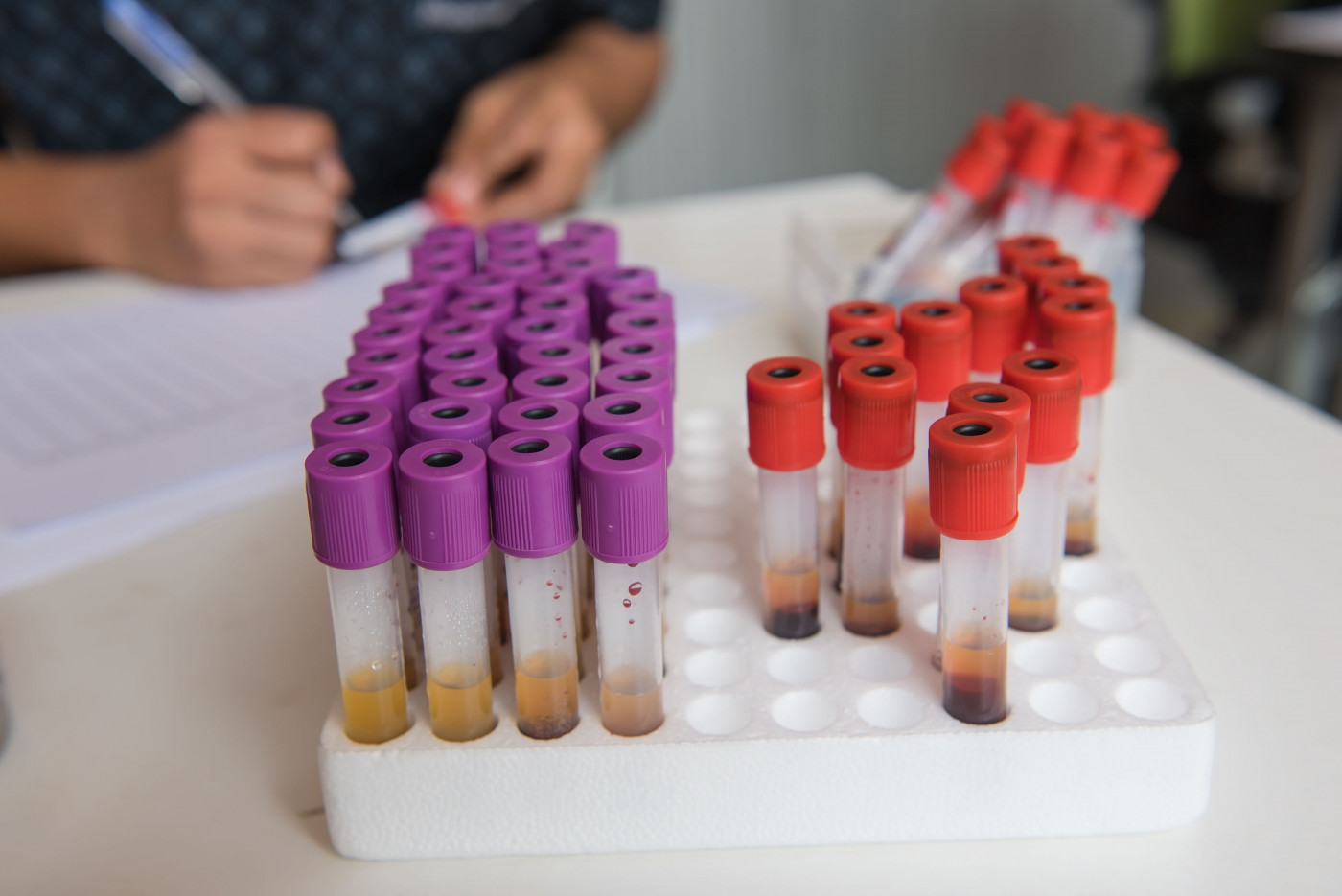
To test the hypothesis, they gathered blood samples from 73 MG patients (mean age 59.1), including 20 who were seronegative for anti-AChR and anti-MuSK antibodies, and 53 who were seropositive for anti-AChR antibodies. The investigators did the same for 49 healthy individuals, who served as controls.
Among the patients, 45 had generalized MG, 24 had ocular MG, and four had an unknown form of the disease.
Analyses showed that MG patients had higher levels of kappa FLC in the blood compared to controls (23.7 mg/L vs. 15.9 mg/L). Higher levels also were seen in seronegative patients (26.8 mg/L) and people with ocular MG (23 mg/L).
In contrast, levels of gamma FLC were not significantly different when comparing MG patients and controls (15.9 mg/L versus 14.5 mg/L).
Additional statistical analyses showed that kappa FLC levels equal or higher than 25 mg/L indicated the presence of MG in seronegative patients and people with ocular MG with a specificity of 98%. Specificity refers to how well a test is able to identify individuals who do not have a disease or particular feature (true negatives).
Importantly, incerased kappa FLC levels found in MG patients were independent of sex, age of disease onset, thymus involvement, or type of treatment received.
“Elevated serum κ [kappa] FLC may serve as a biomarker for MG in suspected patients who are double seronegative and in those with only ocular manifestations when serology is inconclusive,” the investigators concluded.


